
Important: The translations are performed by google. Thank you to forgive the shortcomings that still will understand much of the content of the page. Thank you for your understanding.
The site contains about 1000 pages! Each of them will be translated, which requires a lot of patience and understanding. Thank you very much.
![]()
Our bodies seen to radiography:

The internal frame of the body, the skeleton is a structure whose design is remarkably functional. No skeleton, our body would form a flange and inert mass. To allow a better understanding of this complex, I will present the most known bone, head to toe. Each thumbnail will open the full-size picture. Under each picture, pictures corresponding to key definitions, complement them. But first, as it is a little tradition we start not numbers:
In adults, there are approximately 206 bones, 300 in young children, since some bones are welded during growth. Some people, about 1 in 20, have 13 pairs of ribs instead of 12 for most of us, the lucky ones -)) The average weight of the skeleton is approximately 25% of body weight. The longest bone is THE FEMUR (~ 1/4 size), the larger the BASIN (body width at the hips), and the smallest bone is located in the ear and called STAPEDIEN, it measures only 5 mm long!
SKULL:
face:

1 = Frontal * 2 = Pariétal * 3 = Orbital cavity * 4 = malar bone * 5 = nasal pits * 6 = upper maxilla * 7 = Inferior maxillary * 9 = hole mentonnier
profile:

1 = Frontal * 3 = Orbital cavity* 8 = Pariétal * 10 = occipital * 11 = Temporal * 12 = eyebrow arch * 13 = Bones of the nose * 14 = Mastoïde *
15 = External meatus
Dessus:

1 = Frontal * 8 = Pariétal * 10 = Occipital * 18 = Fronto-parietal suture * 19 = parietal suture or bi-sagittal * 22 = lambdoid suture
Head posterior view:

1 = Pariétal * 2 = Occipital * 3 = Temporal * 4 = Atlas * 5 = Axis * 6 = lower jaw
SPINAL COLUMN

Different postures:
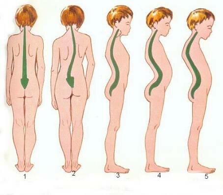
1 = Normal static * 2 = scoliosis * 3 = Normal static * 4 = lordosis * 5 = kyphosis
BUST
Face Anterior: Posterior Face:


Anterior face:
17 = Manubrium (1st Coast) * 18 = Sternum * 19 = ribs * 20 = false ribs * 21 = Floating ribs * 22 = Rachis
Posterior face:
7 = clavicle * 8 = scapula * 17 = Manubrium (1st Coast) * 18 = Ribs * 19 = false ribs * 20 = Floating ribs * 21 = Rachis
ARM AND HAND

9 = Humérus * 10 = Radius * 11 = Cubitus * 12 = Carpes * 13 = metacarpi * 14 = Phalanges * 15 = Phalangines * 16 = Phalangettes
THE BASIN

22 = Rachis * 23 = Iliac bone * 24 = Sacrum * 25 = Coccyx
LEGS AND FEET


legs:
26 = Fémur * 27 = Rotule * 28 = Tibia * 29 = Péroné
feet:
28 = Astragale * 29 = Calcanéum * 30 = Tarses * 31 = Métatarses * 32 = Phalanges * 33 = Phalangines * 34 = Phalangettes
ARTICULATIONS
Different types:

A = Spheroid * B = Ellipsoid : * C = Trochlear * D = Trochoid * E = Sella * F = Plane
Examples of articulations:
Spheroid: Hips and shoulders;
Ellipsoid: Proximal wrist Atlas;
Trochlear: Elbow and knee;
Trochoid: Radius / ulna;
Sella: Thumb;
Plane: plate of the cricoid cartilage, the lower surfaces of the arytenoid cartilages.
A vertebral articulation:

1 = Grand the anterior longitudinal ligament * 2 = Large common posterior vertebral ligament * 3 = yellow ligament * 4 = Ligament on infraspinatus * 5 = Interspinous ligament * 6 = intertransverse ligament * 7 = articular capsule
Bone serves as a lever arm to muscles in contracting, move the framing in the joints. The bones are protective to the brain and spinal cord barrier and shelter in the chest, heart and lungs. Each bone is covered with a layer of osteogenic tissue (which makes the bone), and a very hard and resistant bark: THE PERIOSTEUM. The periosteum contains osteoblasts (see fabrics), able to ensure the replacement of bone ground substance in case of injury.
The durable shell is made of bone Compact bone, while the interior of short bones, flat and epiphyses of long bones, bone or SPONGE Trabecular are met. Trabeculae are disposed along the pressure line and pulling up and a sponge-like or net fabric. A look at the pillars and arches architecture.
THE LONG BONES: (or tubular): Those who compose the ends: humerus, femur, tibia, etc. .. They are recognizable by a central elongate portion or DIAPHYSIS and two joint or epiphyseal ends. Formed between the shaft of compact bone and dug a medullary cavity, and the two formed epiphyseal cancellous bone, are the two metaphyses whose internal structure is intermediate. The marrow cavity contains adult, adipose tissue that forms the bone marrow YELLOW: the femur, humerus, etc..
BONE COURSE: They have two layers of compact bone sandwiching a layer of cancellous bone bones of the skull, or calvaria, vertebrae, tarsus (foot) and carp (hand).
BONE PLATES: scapula, sternum, pelvic, cranial vault, and ribs.
The intertrabecular spaces of short bones, flat, and the epiphyses of long bones contain red marrow. It is in this red marrow, which are formed ERYTHROCYTE or red blood cells, the GRANULOCYTE (a type of white blood cell) and THROMBOCYTES or platelets. During early childhood, the red marrow is present in all bones.
Like the shell of a walnut, the skull is a very hard shell that protects its contents: THE BRAIN. The skull bone 8 is formed arched and which are welded to each other by serrated edges Called: sutures. It is complemented by the 14 bones of the face.
![]()
Emphysema / Cells / Tissues / the Blood / platelets / White Blood Cells
Acne / Muscles / Skeleton / Circulatory Device / Digestive Tract
Breathing Apparatus / Urinary system / Lymphatic System / Immune System
Reproduction & Growth / Special Skin
![]()